Multiphoton Excitation
 Left
Left: A single input photon (blue) excites
a single photon (green).
Right: Multiphoton Excitation.
Two input photons (red) combine to excite
a single photon (green).
MPE is the most simple form of Multiphoton Microscopy (MPM)
which is used to
excite fluorescence. A pulsed laser is used to
provide simultaneous excitation with two (or three)
photons of low energy, exciting the fluorophore to
the same level as one high energy photon. MPE has
many advantages over normal laser scanning
microscopy. The use of lower energy photons
(infrared) reduces damage to the sample and also
permits deeper penetration into scattering samples.
Multiphoton excitation occurs only at the focal
plane, removing the need for pinhole apertures.
As well as excitation of extraneous fluorophores, MPE
can be used to excite indigenous tissue components
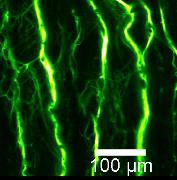
such as NAD(P)H, flavins, and porphyrins. The figure
below shows an MPE image of artery wall
using elastin as the intrinsic fluorophore.
Example

Elastin in an artery wall viewed with Multiphoton Excitation

 Elastin in an artery wall viewed with Multiphoton Excitation