|
| Navigation<> |

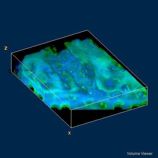
3D reconstruction of a microscopic lesion at the articular surface of cartilage, imaged using SHG (blue) and TPF (green).
The lesion is about 15microns in depth and could be one of the earliest stages of osteoarthritic damage.
The size of the reconstruction is 227 x 164 x 40 microns.