|
| Navigation<> |
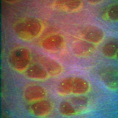
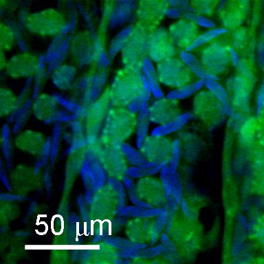
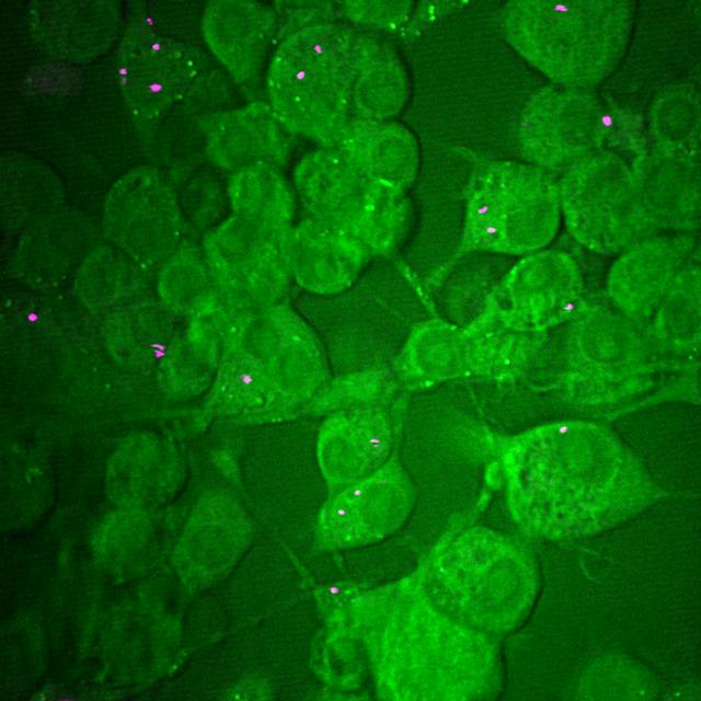
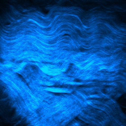
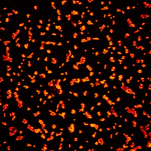
Multi-photon image of articular cartilage in the superficial zones, taken using CARS (red), TPF (green) and SHG (blue).
The size of this image is 135 x 135 microns. The CARS imaging shows the cells within the tissue to be imaged which
would otherwise appear as dark voids. The SHG shows the type II collagen which makes up 20% of the matrix volume
and the TPF image
shows auto-fluorescence from the peri-cellular matrix and from a network of elastin fibres within the sample