|
| Navigation<> |
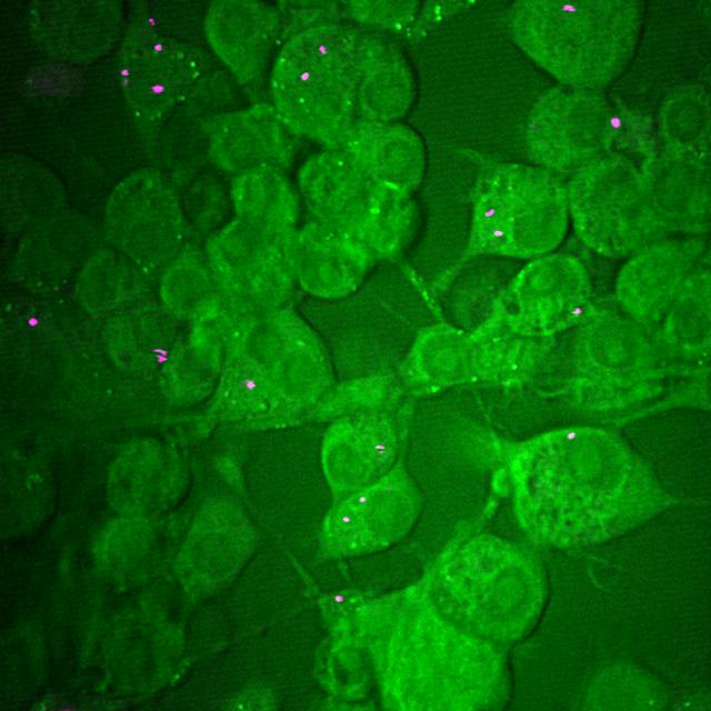
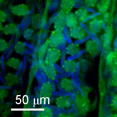
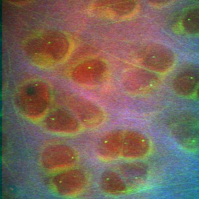
CARS microscopy can be used to visualise polystyrene nanoparticles uptaken by live cells.
The figure shows 100 nm polystyrene beads in cultured macrophage cells; the vibrational signatures of the
nanoparticles and the cells can be separated by tuning (&omega p &omegas).
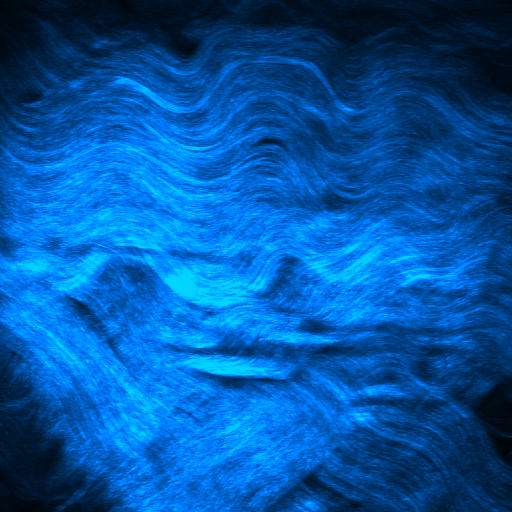
The green image contrast corresponds
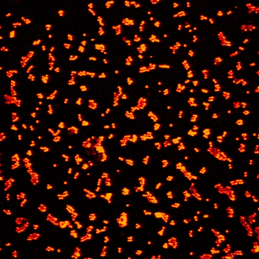
to the CH2 of lipids (2485 cm-1) and the red C=C vibrational stretch of polystyrene (~1600 cm-1).
This study is facilitated by the strong Raman response and high density of
CH2 groups in lipids and C=C in polystyrene, which yield strong CARS signals.